
PB Aptamer Details
ID# 9480
Structure
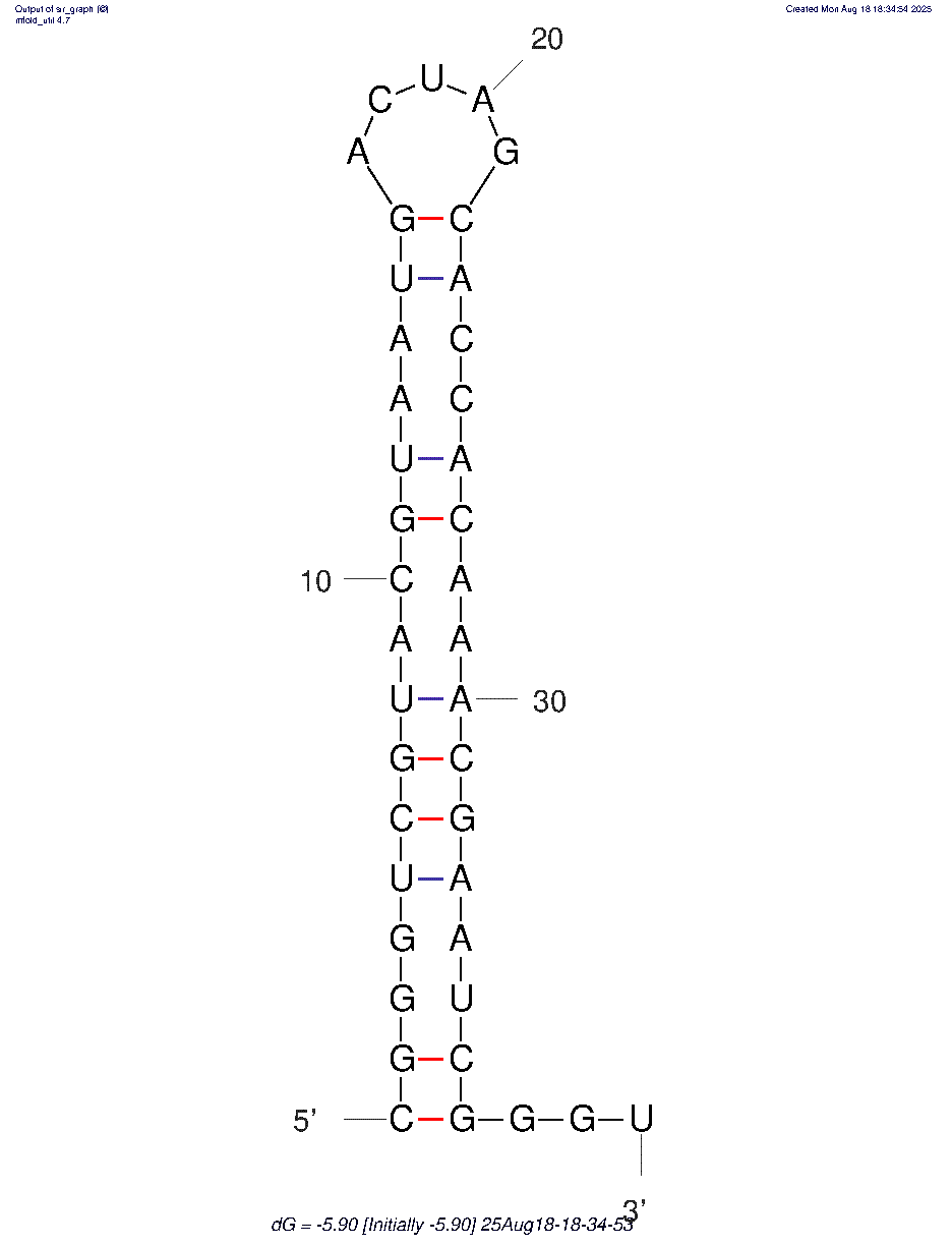
Chemistry
DNA
Target
prostate cancer cells
Category
Cells
Affinity (Kd)
Nude mouse bearing PC3 PBs was chosen to perform aptamer SELEX by a library composed of different ssDNA sequences. When the tumor models were ready about 4–6 weeks after inoculation (nude mouse at the middle stage of PC3 PB development before body paralysis), 10 μg thioaptamer library was intravenously injected to mice. After 4 hours circulation, mice were sacrificed, and bone marrow was collected and frozen in liquid nitrogen. Tissues were homogenized, and DNA was extracted for PCR amplification with aptamer library-specific primers.
Length
30
Aptazyme
No
Sequence (3-char table code)
G
A
C
U/T
dCpdTpdCpdTpdApdTpdTpdGpdApdTpdGpdCpdCpdTpdGpdCpdCpdTpdGpdCpdGpdTpdGpdCpdGpdTpdGpdCpdTpdTpdGpdTpdApdGp
Molecular Weight: 9211 g/mole
Extinction Coefficient: 269900
Binding Conditions / Buffer
71.1 °C
Binding Temp: PCR products were filtered by a Millipore YM-30 filter to purify dsDNA product. The product was incubated with streptavidin-coated magnetic beads in buffer (10 mM Tris–HCl, 2 M NaCl, and 1 mM EDTA [pH 7.5]) for 20 minutes. Biotin on the reverse strand of the dsDNA binds to the streptavidin beads, followed by separation of the two DNA strands in melting solution (0.1 M NaOH). The forward strands were collected after filter with Millipore YM-10 filter for the next round of selection.
Refolding Protocol
Note: Information on this aptamer oligo was obtained from the literature and hasn't been validated by Aptagen.
Reference
Chen L, He W, Jiang H, Wu L, Xiong W, Li B, Zhou Z, Qian Y. In vivo SELEX of bone targeting aptamer in prostate cancer bone metastasis model. Int J Nanomedicine. 2018 Dec 21;14:149-159. doi: 10.2147/IJN.S188003. PMID: 30613143; PMCID: PMC6306056.
Request a Quote
Have your aptamer oligo synthesized by Aptagen.